 乳腺癌的影像学诊断

《乳腺癌的影像学诊断》由会员分享,可在线阅读,更多相关《乳腺癌的影像学诊断(29页珍藏版)》请在装配图网上搜索。
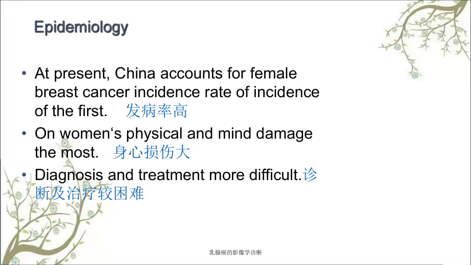
1、乳 腺 癌 的 影 像 学 诊 断 At present, China accounts for female breast cancer incidence rate of incidence of the first. 发 病 率 高 On womens physical and mind damage the most. 身 心 损 伤 大 Diagnosis and treatment more difficult.诊断 及 治 疗 较 困 难 乳 腺 癌 的 影 像 学 诊 断 1. Differentiation of low breast cancer:分 化低 的 乳 腺 癌
2、1) hardware carcinoma硬 癌 2) medullary carcinoma髓 样 癌 3) diffuse carcinoma (also known as inflammatory cancer)弥 漫 性 癌 (亦 称 炎 性 癌 ) 4) mucinous carcinoma (also known as colloid carcinoma) 粘 液 癌 (亦 称 胶 样 癌 ) 乳 腺 癌 的 影 像 学 诊 断 2. Highly differentiated breast cancer:分 化 高 的 乳腺 癌 1) adenocarcinoma腺 癌 2) d
3、uctal carcinoma (also known as tube cancer)导 管 癌 (亦 称 管 内 癌 ) 3)papillary carcinoma (also known as papillary adenocarcinoma)导 管 癌 (亦 称 管 内 癌 ) 4) eczema-like cancer (also known as Pagets nipple disease)湿 疹 样 癌 (亦 称 Paget氏 乳 头 病 ) 乳 腺 癌 的 影 像 学 诊 断 Breast lumps 乳 房 肿 块 Nipple discharge 乳 头 溢 液 Change
4、s in the breast skin and contour乳房 皮 肤 及 轮 廓 改 变 乳 腺 癌 的 影 像 学 诊 断 Ultrasound Mammography 钼 靶 X线 MRI Near infrared scanning 近 红 外 线 扫 描 Positron emission tomography (PET) Enhanced CT 乳 腺 癌 的 影 像 学 诊 断 Solid hypoecho低 回 声Without capsule无 包 膜 Border clearance边 界 清 Heterogeneous不 均 匀internal echo Stron
5、g echo can be seen clustered calcifications钙 化 乳 腺 癌 的 影 像 学 诊 断 See the upper outer quadrant mass in shallow leaf浅 分 叶 肿 块 影 Less clear boundary See clusters of calcification 其 内 见 成 簇钙 化 乳 腺 癌 的 影 像 学 诊 断 MRI should no longer be regarded as an adjunct to mammography but as a distinct breast cancer
6、 in its earliest stage. “MRI 不 再 是 钼 靶 X线 的 辅 助 方 法 , 而 是作 为 乳 腺 癌 早 期 诊 断 的 独 立 检 查 方 法 。 ” 乳 腺 癌 的 影 像 学 诊 断 T1WI low signal T2WI Increased signal 乳 腺 癌 的 影 像 学 诊 断 Irregular不 规 则 mass Lobulated分 叶 状 edges Blur模 糊 , rough毛 糙 Crab-like growth呈蟹 足 样 生 长 Ill-defined, 乳 腺 癌 的 影 像 学 诊 断 Liquefaction ne
7、crosis液 化 坏 死 seen in the larger lesions 乳 腺 癌 的 影 像 学 诊 断 Quick significantly明 显 enhanced Rapid clearance廓 清 Around the beginning of the spread of the disease centers 向 心 性 强 化 Liquefaction necrosis and bleeding液 化 坏 死及 出 血 has always been part of no enhancement 乳 腺 癌 的 影 像 学 诊 断 Type Clearance typ
8、e type 持 续 型 平 台 型 廓 清 型Persistent Platform Type 乳 腺 癌 的 影 像 学 诊 断 Most of the benign lesions showed persistent持 续 型 to enhance Most malignant lesions showed clearance-type廓 清 型 enhanced Platform-type平 台 型 can be benign lesions, but also for malignant lesions. 乳 腺 癌 的 影 像 学 诊 断 Level 0: assessed评 估
9、is not complete, to further assess the need for other imaging Level 1: no abnormal findings无 异 常 发 现 Level 2: Benign found良 性 发 现 - routine follow-up Level 3: Possible positive findings可 能 良 性 发 现 - short-term follow-up Level 4: Low - Moderate suspicious malignant低 -中 度 可疑 恶 性 - consider biopsy Leve
10、l 5: highly suspected malignancy高 度 怀 疑 恶 性 - Level 6: malignant lesions biopsy confirm 已 活 检 证 实 的恶 性 病 变 乳 腺 癌 的 影 像 学 诊 断 Breast fibroadenoma: 纤 维 腺 瘤 Often occurs in young female, Mass often smooth光 滑 edge, Similar density and glands腺 体 , Coarse calcification within the visible, Strengthen even,
11、 slow, was eccentric. 乳 腺 癌 的 影 像 学 诊 断 Hyperplasia增 生 : Local gland increased, edges blur模 糊 Enhanced with a slow, progressive渐 进 性enhancement, In the late enhancement, lesion degree of enhancement increasing trend 乳 腺 癌 的 影 像 学 诊 断 Breast cyst囊 肿 : appear as significant long T1 and long T2 singal
12、intensity 乳 腺 癌 的 影 像 学 诊 断 纤维腺瘤1a) show low T1WI signal mass at the left breast1b) show high T2WI signal mass, edge clear 乳 腺 癌 的 影 像 学 诊 断 纤维腺瘤 2a) T2WI mass signal was slightly lower2b) Enhanced scan mass lesion in the breast deep, sharp edges and strengthen a more even 乳 腺 癌 的 影 像 学 诊 断 纤维腺瘤 3a)
13、 Enhanced mass in the right breast showed a more homogeneous均 匀 enhancement3b) Curve for the lesion type type curve for the normal glands 乳 腺 癌 的 影 像 学 诊 断 浸润性导管癌4a) Mass within the left breast was significantly明 显enhanced 4b) Dynamic enhancement curves of the type (clearance type) 乳 腺 癌 的 影 像 学 诊 断
14、 浸润性导管癌5a) X-ray image Right breast near the chest wall see the mass5b) Significantly enhanced lesions, irregular 不 规则 margin 乳 腺 癌 的 影 像 学 诊 断 浸润性导管癌6a) X-ray image Large calcified lesions in the breast 6b) The outer upper quadrant lesions within the chip heterogeneous 不 均 匀 enhancement 乳 腺 癌 的 影 像
15、 学 诊 断 乳腺囊肿 乳 腺 癌 的 影 像 学 诊 断 乳腺增生 乳 腺 癌 的 影 像 学 诊 断 Middle-aged women with multiple多 发 Was infiltrating浸 润 the surrounding tissue invasion aptitude Mass of edge the rough rf毛 糙 Enhance the rapid clearance was quickly apparent Strengthen the Mind Mass examination hard, poor activity活动 度 差 乳 腺 癌 的 影 像 学 诊 断 乳 腺 癌 的 影 像 学 诊 断
- 温馨提示:
1: 本站所有资源如无特殊说明,都需要本地电脑安装OFFICE2007和PDF阅读器。图纸软件为CAD,CAXA,PROE,UG,SolidWorks等.压缩文件请下载最新的WinRAR软件解压。
2: 本站的文档不包含任何第三方提供的附件图纸等,如果需要附件,请联系上传者。文件的所有权益归上传用户所有。
3.本站RAR压缩包中若带图纸,网页内容里面会有图纸预览,若没有图纸预览就没有图纸。
4. 未经权益所有人同意不得将文件中的内容挪作商业或盈利用途。
5. 装配图网仅提供信息存储空间,仅对用户上传内容的表现方式做保护处理,对用户上传分享的文档内容本身不做任何修改或编辑,并不能对任何下载内容负责。
6. 下载文件中如有侵权或不适当内容,请与我们联系,我们立即纠正。
7. 本站不保证下载资源的准确性、安全性和完整性, 同时也不承担用户因使用这些下载资源对自己和他人造成任何形式的伤害或损失。
