 外文翻译--Bioinformatic Analysis of UL27 gene of Duck Plague Virus CHv Strain


《外文翻译--Bioinformatic Analysis of UL27 gene of Duck Plague Virus CHv Strain》由会员分享,可在线阅读,更多相关《外文翻译--Bioinformatic Analysis of UL27 gene of Duck Plague Virus CHv Strain(6页珍藏版)》请在装配图网上搜索。
1、Bioinformatic Analysis of UL27 gene of Duck Plague Virus CHv Strain Long Jiang,Dan Lin,Anchun Cheng,Mingshu Wang,Dekang Zhu,Xiaoyue Chen Avian Disease Research Center College of Veterinary Medicine of Sichuan Agricultural University,Yaan,Sichuan,625014,China Anchun Cheng,Xiaoyue Chen Epizootic Disea
2、ses Institute of Sichuan Agricultural University,Yaan,Sichuan,625014,China E-mail: Anchun Cheng,Mingshu Wang,Dekang Zhu,Renyong Jia,Qihui Luo,Hengmin Cui,Yi Zhou,Yin Wang,Zhiwen Xu,Zhengli Chen,Xiaoyue Chen,Xiaoyu Wang Key Laboratory of animal diseases and Human Health of Sichuan Province,Sichuan Ag
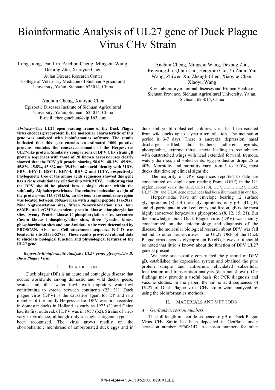
3、ricultural University,Yaan,Sichuan,625014,China AbstractThe UL27 open reading frame of the Duck Plague virus encodes glycoprotein B,the molecular characteristic of this gene was analyzed with bioinformatics software.The results indicated that this gene encodes an estimated 1000 putative proteins,con
4、tains the conserved domain of the Herpesvirus UL27-like protein.Similarity comparisons of DPV CHv strain gB protein sequences with those of 20 known herpesviruses clearly showed that the DPV gB protein sharing 58.0%,48.1%,45.9%,45.8%,45.8%,45.8%and 39.4%amino acid identity with MDV,PRV,EFV-1,HSV-1,E
5、HV-4,BHV-2 and ILTV,respectively.Phylogenetic tree of the amino acids sequences showed this gene has a close evolutionary relationship with MDV,indicating that the DPV should be placed into a single cluster within the subfamily Alphaherpesvirinae.The relative molecular weight of the protein was 113.
6、8781kDa,an obvious transmembrane region was located between 860aa-883aa with a signal peptide 1aa-20aa.Nine N-glycosylation sites,fifteen N-myristoylation sites,four cAMP-and cGMP-dependent protein kinase phosphorylation sites,twenty Protein kinase C phosphorylation sites,seventeen Casein kinasephos
7、phorylation sites,three Tyrosine kinase phosphorylation sites and one Amidation site were determined by PROSCAN.Also,one Cell attachment sequence R-G-D was located in site 525aa-527aa.These results provided rational data to elucidate biological function and physiological features of the UL27 gene.Ke
8、ywords-Bioinformatic Analysis;UL27 gene;glycoprotein B;Duck Plague Virus I.INTRODUCTION Duck plague(DP)is an acute and contagious disease that occurs worldwide among domestic and wild ducks,geese,swans,and other water fowl,with migratory waterfowl contributing to spread between continents(23,31).Duc
9、k plague virus(DPV)is the causative agent for DP and is a member of the family Herpesviridae.DPV was first recorded in domestic ducks in Holland as early as 1923(1)and China had its first outbreak of DPV was in 1957(32).Strains of virus vary in virulence,although only a single antigenic type has bee
10、n recognized.The virus grows readily on the chorioallantoic membrane of embryonated duck eggs and in duck embryo fibroblast cell cultures,virus has been isolated from wild ducks up to a year after infection.The incubation period is 3-7 days.There is anerexia,depression,nasal discharge,ruffled,dull f
11、eathers,adherent eyelids,photophobia,extreme thirst,ataxia leading to recumbency with outstretched wings with head extended forward,tremors,watery diarrhea,and soiled vents.Egg production drops 25 to 40%.Morbidity and mortality vary from 5 to 100%,most ducks that develop clinical signs die.The major
12、ity of DPV sequences reported to date are concentrated on single open reading frame(ORF)in the UL region,recent years,the UL2,UL6(30),UL7,UL11,UL27,UL32,UL35(20)and UL36 gene sequences had been illuminated in our lab.Herpesviridae have an envelope bearing 12 surface glycoproteins(8).Of these glycopr
13、oteins,only gB,gD,gH,and gL participate in viral cell entry and fusion,gB is the most highly conserved herpesvirus glycoprotein(8,12,15,21).But the knowledge about Duck Plague virus(DPV)was mainly concentrated on the epidemiology and diagnosis of the disease,the molecular biological research about D
14、PV was fall behind to other herpesviruses.The UL27 ORF of the Duck Plague virus encodes glycoprotein B(gB),however,it should be noted that little is known about the function of DPV UL27 gene at present.We have successfully constructed the plasmid of DPV gB,established the expression system and obtai
15、ned the pure protein sample and antiserum,elucidated subcellular localization and transcription analysis(data not shown).Our findings may provide a useful basis for PCR diagnosis and vaccine studies.In the paper,the amino acid sequences of UL27 of Duck Plague virus CHv strain were analyzed by using
16、the bioinformatics methods.II.MATERIALS AND METHODS A.GenBank accession numbers The full length nucleotide sequence of gB of Duck Plague Virus CHv Strain has been deposited in GenBank under accession number EF608147.Accession numbers for other 978-1-4244-4713-8/10/$25.00 2010 IEEEmembers of the Herp
17、esviridae sequences used in this study were shown in Table 1.B.Bioinformatic analysis of DPV gB The raw data were edited using the EditSeq program in the DNAStar software package(Lasergene).Multiple alignments,sequence similarity and phylogenetic analyses of nucleotide sequences between Tianjin stra
18、in and 20 known herpesviruses were performed with the DNAStar MegAlign program(version 5.00)using the ClustalW method.Table 1 Accession No.of gB of Herpesviridae Prediction of Signal peptide and transmembrane segment were analyzed with SignalP 3.0 Server(http:/www.cbs.dtu.dk/services/SignalP/)and TM
19、HMM Server v.2.0(http:/www.cbs.dtu.dk/services/TMHMM/),respectively.In this paper,we also use a noval web tool Phobius(http:/www.ebi.ac.uk/Tools/phobius/)to predict signal peptide and transmembrane segment as contrast.Primary and Secondary structure were examined by ProtParam on the Expasy website(h
20、ttp:/www.expasy.org)and SOPM(Geourjon and Delage,1994)(http:/npsa-pbil.ibcp.fr/).The three-dimensional model of gB was generated on the Swiss-Model server(http:/swissmodel.expasy.org/).Functional sites were predicted by PROSCAN against PROSITE database(http:/npsa-pbil.ibcp.fr/).Antigenic determinant
21、s was analyzed on the Mobyle server(http:/mobyle.pasteur.fr/cgi-bin/portal.py).III.RESULTS A.Similarity comparison Similarity comparison of DPV CHv strain gB nucleotide sequences with those of 20 known herpesviruses clearly showed that the DPV gB protein sharing 58.0%,48.1%,45.9%,45.8%,45.8%,45.8%an
22、d 39.4%amino acid identity with MDV,PRV,EFV-1,HSV-1,EHV-4,BHV-2 and ILTV,respectively,and it was closest genetically with MDV(58%)(Table 2.).B.Phylogenetic analysis Phylogenetic analysis based on nucleotide sequences of gB of the DPV CHv strain and 20 strains of Herpesviridae clearly showed that the
23、 viruses were roughly divided into three phylogenetic clusters,including Alphaherpesvirinae,betaherpesvirinae and Gammaherpesvirinae.Phylogenetic analysis demonstrated that DPV gB protein had more homologous with gB protein of alphaherpesvirus subfamily than with those of beta-and gamma-subfamily,so
24、 it should be assigned to the subfamily Alphaherpesvirinae.The DPV CHv strain formed a single cluster with MDV,and was most closely related to MDV.Phylogeny trees illustrating the relationships observed are shown in Fig.1.Table 2 Similarity analysis of the nucleotide sequences of gB gene between DPV
25、 CHv strain and other herpesviruses(%)Figure 1 The Phylogeny tree based on the complete gB nucleotide sequences of DPV CHv strain and other herpesviruses C.Signal peptide and transmembrane domain analysis Using SignalP 3.0 and TMHMM v.2.0,most likely signal peptide cleavage site were found between p
26、os.20aa and 21aa(Fig.2)and the prodicted transmembrane domain of gB protein may span amino acids 861-883(Fig.3).Phobius is a web tool to predict transmembrane topology of protein with a hidden Markov mode,it had predicted the transmembrane domain of gB protein to span amino acids 860-883,and ectodom
27、ain span amino acids 1-859,cytoplasmic span amino acids 884-1000 were all identified,but no signal peptide was found by the tool(Fig.4).Viruses Accession No.Duck enteritis virus EF608147 Mareks disease herpesvirus(strain RB-1b)P18538 Human herpesvirus 3 strain Dumas P09257 Bovine herpesvirus type 1.
28、1(strain Cooper)P12640 Equine herpesvirus 1(strain V592)Q6S6T9 Suid herpesvirus 1(strain Indiana-Funkhauser/Becker)P08355 Equine herpesvirus type 4(strain 1942)P17472 Ateline herpesvirus 1 Q04463 Human herpesvirus 2(strain 333)P06763 Saimiriine herpesvirus 1 Q04464 Bovine herpesvirus type 2(strain B
29、MV)P12641 Infectious laryngotracheitis virus(strain SA-2)P27415 Human herpesvirus 1(strain KOS)P06437 Epstein-barr virus(strain ag876)P0C763 Human herpesvirus 5(strain AD169)P06473 Rhesus cytomegalovirus(strain 68-1)P89053 Murine cytomegalovirus(strain Smith)P27171 Guinea pig cytomegalovirus(strain
30、22122/ATCC VR682)(GPCMV)Q69024 Human herpesvirus 6 strain Z29 P36320 Human herpesvirus 7 strain JI P52352 Tupaiid herpesvirus 2 Q9WRL5 D.Primary structure analysis ExPASy Proteomics tools were used to predict physical and chemical characteristics of DPV gB.The results showed that DPV gB protein has
31、1000aa with a molecular weight of 113.8781 kDa,Isoelectric point(pI)8.87,Aliphatic index:76.24,C5011H7887N1431O1516S45 formula,and grand average of hydropathicity(GRAVY)is-0.478.The total number of negatively charged residues(Asp+Glu)and positive charged residues(Arg+Lys)were 123 and 137,respectivel
32、y.The estimated half-life in mammalian reticulocytes(in vitro),yeast(in vitro)and Escherichia coli(in vitro)were 30,20 and 10 hours,respectively.The instability index(II)is computed to be 42.19,this classifies the protein as unstable.The contents of Arg,Leu,Ser,Val and Ala were 8.9%,8.0%,7.6%,7.5%an
33、d 7.3%,respectively.Fig.2 Predict of signal peptide in gB protein,“C score”indicates the cleavage site probability;“S score”indicates the possible region of signal peptide;“Y score”is a derivative of the C-score,indicates the cleavage site probability.Fig.3 Transmembrane segment profile of gB protei
34、n E.Secondary structure analysis Secondary structure analyzed by SOPM methods showed that the Alpha helix(Hh),Extended strand(Ee),Beta turn(Tt)and Random coil(Cc)occupied 39.9%(399aa),17.8%(178aa),9.1%(91aa)and 33.2%(332aa),respectively(Fig.5,Fig.6).F.Three-dimensional model analysis The crystal str
35、ucture of DPV gB protein(rang 151aa-808aa)was based on template of HSV-1 extracellular domain,and gB protein sequences of DPV and HSV-1 share 47.344%identity(Fig.7).The crystal structure of the HSV-1 gB protein ectodomain contains three protomers,crystallo-Fig.4 Phobius results of transmembrane doma
36、in and signal peptide of gB protein 10 20 30 40 50|MYRRTICYLRDRMPAYFCNSSGPEWRNNPRDVGCDRQGRLLYALYGASTT hhhhhhhhhhtccceeeecccccccccccccccccttcheeeeeeccccc GSRNGGACTTAVRRKEFDRMNRARRVSALHLTAPRRSRFVLSLIMVSVLF ccccccceeeehhhhhhhhhhhhhhhhheeeccttccheeeehhhhheec RPIQPQPIVNATDRPHGLMNDQDTHLDGERLQRGKLSARELIRGV
37、QSTRE ccccccceeeccccccccccccccccthhhhcttccchhhhhhhhhcccc EDKFYVCPPPTGTTVLRFEPSRKCPEAYKGRNWTEGVAVIFKENINPLKF cceeeeccccccceeeeecccccccccccccccctteeeeeettccccch KAMIYYKNVIQTTTWSGTWYKQITNRYTDRVPVALDEITDRIDRFGKCLS heeeeehhheeeeccttcchhhhhhtcccccchhhhhhhhhhhhhhhhhh AAKYLRNNVYVDAFDRDESAKEVPLLASRFNTPDSRAWH
38、TTNDTYTVFGS hhhhhttteeeehcccccchhccheehhcccccccceeeectteeeeecc PWIYRTSTSVNCIVEEVMARSAFPYDYFALANGDVVNMSPFYGYGPTESR ceeecccchhhhhhhhhhhttccccceeeecttceeeecccttccccccc EHNSYSSNRYKQLDGYKVMDLDTHRLASPIKRNFLTTEHYTVGWDWMPKR cccccccccccccttceeeecctthcccccchhheeetteeecccccccc ERVCSMAKWQEVNEMMRAEYGTRYRFASKTLSA
39、SFISNMTQFNLDRLYLG hhhhhhhhhhhhhhhhhhhttcceeecttccchhhhhhhhhccccheehh DCVKRDSEEAIKRIFEEQYASTHVKAGAIEYYLTSGGFILAYQPVVSNTL hhhccchhhhhhhhhhhhhhhhhhhtthheeeecttceeeeecchhhhhh VKMYVQELTRDNRTELALDLLGAMRGDKTREDGRSRHARSVDGGPDGLIR hhhhhhhhccccchhhhhhhhhhhttcccccttccccceeetccccceee GARNIDPYKNYTVRSARVARNADPYKN
40、TTVVKSVMSAQFAMLQYTYDHIQ tcccccttcceeeehhhhhttccccccceehhhhhhhhhhhhhhhhhhhh AHVNDMLSRIAVSWCELQNKESVLWAEMRKVNPSLIASTTVGRRVGARML hhhhhhhhhhhhhhhhhttthhhhhhhhhhccttheecccccchhhhhhh GDVLAVSSCIEVPSSQISLSNSMRIPGGSSCYSRPPVTFSYEKDGGTIDG hhhheecceeeccccceeeccccccccccccccccceeeeeccttcceee MLGENNELLLTREYKEPCAAN
41、HKRYFMFGPNYVLYEEYQFVRQVEAADIQ eettttheeehhhcccccccccceeeeectteeeehhhhhhhhhhhhhhh MVSTFVELNLTMLEDREILPLQVYTREEIRDSGVLDYAEVQRRNQLHSLR hhhhhhhhhhhhhctttccceeeechhhhcttceeehhhhhhttthhhhe FYDIDKVIDIDSNYAFMADLTNFFKGLGEAGQAIGKVVVGVAGAVVSTVS eeccceeeehccchhhhhhhhhhhhhhhhtthhhteeeeeettheeeeht GISSLLSNPFGALAI
42、GLIVVAGLVVAFLAYRYISKLRNDPVKTLYPMTMK thhhhhccccchhhhheeeetthhhhhhhhhhhhhccccccceechhhhh TLKNEAKLTPRGEADGEDEDEFDESKLEQAREMVRYMALLSASERTEKKA hhhtttcccccccccccchhhhhhhhhhhhhhhhhhhhhhhhhhhhhhhh RKKNSRTALLSNHLSNLRSRSNGKKYSKVEDEYEDGDSADETEILVTDRV hccccchhhhhhhhhhhhtttttcccchhhhhhcttcccthhheeeeccc Fig.5 pre
43、diction of secondary structure of DPV gB protein,The letters h,e,t and c in the figure represent the structures of alpha helix,beta turn,extended strand and random coil,respectively.Fig.6 Analysis of distribution of secondary structure in gB protein graphical suggests that HSV-1 gB may exist as a tr
44、imer which with unexpected homology to glycoprotein G from vesicular stomatitis virus(VSV G)(7,25)and gB from EpsteinBarr virus(EBV)(18).Each protomer can be divided into five distinct regions or domains,and each extends a C-terminal arm from one end of the molecule to the other,inserting it into th
45、e junction between the other two protomers,multiple contacts between protomers throughout the molecule contribute to trimer stability.G.Functional sites analysis Nine N-glycosylation sites,fifteen N-myristoylation sites,four cAMP-and cGMP-dependent protein kinase phosphorylation sites,twenty Protein
46、 kinase C phosphorylation sites,seventeen Casein kinasephosphorylation sites,three Tyrosine kinase phosphorylation sites and one Amidation site were determined by PROSCAN.Also,one cell attachment sequence R-G-D(Arg-Gly-Asp)was located in site 525aa-527aa of gB protein.H.Antigenic determinants analys
47、is The length of these antigenic determinants rang from 6aa to 52aa(Table.3).The sequence of highest score was located between 834aa-885aa,containing the predicted transmembrane domain.All antigenic determinants but 936aa-942aa and 957aa-967aa were located in ectodomain.The predicted cell attachment
48、 sequence R-G-D was not included in any of these antigenic determinants.IV.DISCUSSION To date,the Herpesviridae has been divided into three subfamilies,the Alpha-,Beta-,and Gammaherpesvirinae,but which subfamily especially genus was DPV assigned in was also argumentative.DEV was classified as unassi
49、gned virus in the family Herpesviridae according to the Eighth International Committee on Taxonomy of Viruses(ICTV)although it was previously grouped in the subfamily Alphaherpesvirinae(26),so the molecular biology data such as the distinct biological properties of a single gene or its genomic attri
50、butes can provide a reliable reference.DPV gB protein plays a dominant role in viral cell entry and fusion,and gB is the most highly conserved herpesvirus glycoprotein.The gB protein trees showing that the CHv strain was positioned in the subfamily Alphaherpesvirinae and was most closely related to
51、MDV.Fig.7 A 3D model of cytoplasmic domain of gB protein.(http:/swissmodel.expasy.org/)Table.3 Analysis of distribution of antigenic determinants No.ScoreLengthStart Position Sequence End position Max score position 11.24652834IGKVVVGVAGAVVSTVSGISSLLSNPFGALAIGLIVVAGLVVAFLAYRYISK 885872(G)21.21721650
52、LGDVLAVSSCIEVPSSQISLS 670656(V)31.18939470ASTHVKAGAIEYYLTSGGFILAYQPVVSNTLVKMYVQEL 508493(Q)41.1829768ILPLQVYTR 776771(L)51.1792587SRFVLSLIMVSVLFRPIQPQPIVNA 11193(L)61.1778153KFYVCPPP 160157(C)71.17112444LDRLYLGDCVKR 455450(G)81.1479309SVNCIVEEV 317311(N)91.1441137QGRLLYALYGA 4743(A)101.1391075RVSALH
53、LTAP 8479(L)111.13813678GSSCYSRPPVTFS 690684(R)121.13710781DSGVLDYAEV 790787(Y)131.13311577NTTVVKSVMSA 587583(S)141.13218730PNYVLYEEYQFVRQVEAA 747741(V)151.1288271KEVPLLAS 278276(L)161.12810245FGKCLSAAKY 254251(A)171.1268229DRVPVALD 236232(P)181.12613749IQMVSTFVELNLT 761755(F)191.1257186GVAVIFK 1921
54、89(V)201.12411608SRIAVSWCELQ 618614(W)211.1169256RNNVYVDAF 264262(D)221.1157936YMALLSA 942937(M)231.1157516LALDLLGA 523519(D)241.1067796LHSLRFY 802799(L)251.10216197PLKFKAMIYYKNVIQT 212207(K)261.10111957TALLSNHLSNL 967961(S)271.1009632NPSLIASTT 640634(S)281.09917589FAMLQYTYDHIQAHVND 605600(Q)291.094
55、9322AFPYDYFAL 330327(Y)301.09310560NYTVRSARVA 569566(A)311.09074RTICYLR 106(I)321.08510426FASKTLSASF 435434(S)331.083715AYFCNSS 2117(F)341.0787715KEPCAAN 721717(P)351.0786707ELLLTR 712711(T)361.0758162GTTVLRFE 169168(F)371.0747373THRLASP 379377(A)381.0747401ERVCSMA 407406(M)391.0737804IDKVIDI 810805
56、(D)401.07110295YTVFGSPWIY 304298(F)411.0597171SRKCPEA 177176(E)421.0557620KESVLWA 626623(V)431.0396387TEHYTV 392387(T)Homologues of gB may possess common structural and functional domains,complementation experiments showed that the gB glycoprotein of HSV-1 could be substituted with the corresponding
57、 homologue from either PRV(27)or Bovine herpesvirus 1(BHV-1)(29).Also,gB-null PRV could be complemented by gB protein of BHV-1(2).When using conventional transmembrane topology and signal peptide predictors,such as TMHMM and SignalP,there is a substantial overlap between these two types of predictio
58、ns.This impairs predictions of 5-10%of the proteome,hence this is an important issue in protein annotation.To address this problem,Kll L designed a hidden Markov model,Phobius(17),that combines transmembrane topology and signal peptide predictions.The method makes an optimal choice between transmemb
59、rane segments and signal peptides,and also allows constrained and homology-enriched predictions.It had predicted the transmembrane domain of gB protein to span amino acids 860-883,but no signal peptide was found by the tool.Comparative structure analyses of DPV gB and HSV-1 gB,VSV G,may shed light o
60、n gB residues that may be involved in conformational changes and membrane fusion.Also,the DPV gB struture reveals that,despite the high sequence conservation of gB in herpesviruses,the relative orientations of individual domains,the surface charge distributions,and the structural details of DPV gB m
61、ay differ from the HSV-1 protein and VSV G,indicating regions and residues that may have important roles in virus-specific entry.DPV is composed of a linear,double stranded DNA genome with 64.3%G+C content,higher than any other reported avian Alphaherpesvirinae(22).DPV gB has 1000aa with a molecular
62、 weight of 113.8781 kDa,the assembly and processing of glycoprotein B is still unknown.Infectious laryngotracheitis virus(ILTV)gB is initially synthesized as a 110 kDa monomeric precursor protein in the endoplasmic reticulum(ER),rapidly assembles into homodimers composed of 100 kDa subunits,and then
63、 transported to the Golgi apparatus where it is rapidly cleaved to form two disulphide-linked species of 58 kDa.The apparent reduction in mass(from 110 to 100 kDa)of the mature form of gB during processing in the Golgi apparatus appears to be a common feature of avian herpesvirus gB proteins.The gB
64、homologue of ILTV is processed in a manner similar to the gB homologues of Mareks disease virus(MDV),varicella-zoster virus(VZV)(6),bovine herpesvirus 1(BHV-1)(2)and human cytomegalovirus(CMV)(28).The cell attachment sequence R-G-D,found in fibronectin,is crucial for its interaction with its cell su
65、rface receptor,an integrin(10),this is also true for HHV-8 gB could mediate cell adhesion via its RGD motif interaction with the cell surface integrin molecules(11,24).What has been called the“R-G-D”tripeptide is also found in the sequences of a number of other proteins,where it has been shown to pl
66、ay a role in cell adhesion(5,13,14,16,19).These proteins are some forms of collagens,fibrinogen,vitronectin,von Willebrand factor(VWF),snake disintegrins,and slime mold discoidins.The structure of HSV-1 gB can taken to represent the structure of all herpesvirus gB proteins,and in analyzing properties such as antigenicity,we can map results from various herpesviruses onto it(7).Herpesvirus gB glycoprotein is incorporated into membranes and appears on the surface of both virions and infected cells
- 温馨提示:
1: 本站所有资源如无特殊说明,都需要本地电脑安装OFFICE2007和PDF阅读器。图纸软件为CAD,CAXA,PROE,UG,SolidWorks等.压缩文件请下载最新的WinRAR软件解压。
2: 本站的文档不包含任何第三方提供的附件图纸等,如果需要附件,请联系上传者。文件的所有权益归上传用户所有。
3.本站RAR压缩包中若带图纸,网页内容里面会有图纸预览,若没有图纸预览就没有图纸。
4. 未经权益所有人同意不得将文件中的内容挪作商业或盈利用途。
5. 装配图网仅提供信息存储空间,仅对用户上传内容的表现方式做保护处理,对用户上传分享的文档内容本身不做任何修改或编辑,并不能对任何下载内容负责。
6. 下载文件中如有侵权或不适当内容,请与我们联系,我们立即纠正。
7. 本站不保证下载资源的准确性、安全性和完整性, 同时也不承担用户因使用这些下载资源对自己和他人造成任何形式的伤害或损失。
