 足舟骨背侧入路

《足舟骨背侧入路》由会员分享,可在线阅读,更多相关《足舟骨背侧入路(8页珍藏版)》请在装配图网上搜索。
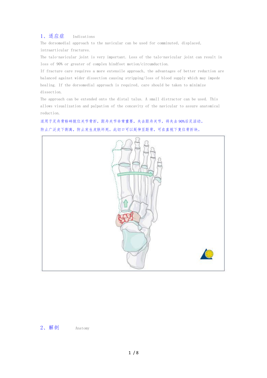
1、1、适应症 IndicationsThe dorsomedial approach to the navicular can be used for comminuted, displaced, intraarticular fractures.The talo-navicular joint is very important. Loss of the talo-navicular joint can result in loss of 90% or greater of complex hindfoot motion/circumduction.If fracture care requi
2、res a more extensile approach, the advantages of better reduction are balanced against wider dissection causing stripping/loss of blood supply which may impede healing. If the dorsomedial approach is required, care should be taken to minimize dissection.The approach can be extended onto the distal t
3、alus. A small distractor can be used. This allows visualization and palpation of the concavity of the navicular to assure anatomical reduction.适用于足舟骨粉碎脱位关节骨折,距舟关节非常重要,失去距舟关节,将失去90%后足活动。防止广泛皮下剥离,防止发生皮肤坏死。此切口可以延伸至距骨,可在直视下复位骨折块。2、解剖Anatomy The dorsomedial approach to the navicular is made between the t
4、ibialis anterior tendon and extensor hallucis longus (EHL) tendon. The approach should be made straight down from skin to periosteum without raising flaps or any unnecessary dissection. A small distractor can be used to aid in medial column alignment/length and to allow visualization and palpation o
5、f the talonavicular joint. Once reconstruction, bone grafting (if needed) and provisional fixation are accomplished, a dorsal plate can be applied. If needed, the navicular reconstruction can include bridge plating and screw fixation into the cuneiforms. 背侧切口位于胫骨前肌和拇长伸肌腱之间,直接经皮切开至骨膜,不要别离皮瓣。 3、皮肤切口Sk
6、in incisionThe incision uses the interval between the tibialis anterior and the EHL, roughly directly over the fracture.背侧切口位于胫骨前肌和拇长伸肌腱之间,舟骨上方。The incision can be extended distally if incorporation of the cuneiforms into the construct is needed.The incision can be extended proximally to allow inspe
7、ction and palpation of the talonavicular joint.切口可以向远端延伸,如果楔骨有骨折的话,也可向近端延伸探查距舟关节。4、深层别离Deep dissectionOnce down to the periosteum/joint capsule, the tibialis anterior can be retracted medially and the EHL can be retracted laterally. This will expose the dorsum of the navicular.切开至骨膜或关节囊后,将胫前肌向侧牵开,将拇
8、长伸肌腱向外侧牵开,显露舟骨背侧。5、直视关节Visualization of the jointIn a high-energy injury, the comminution may be severe. Care should be taken not to strip the periosteum or joint capsule from any small pieces. If a piece is attached to a proximal piece of joint capsule, then the best course of action may be to flip
9、 it proximally so as not to disrupt its soft-tissue attachments. Once the joint is reconstructed, this “trap-door piece can be reduced and fixed.在高能量损伤,骨折粉碎严重,注意不要从骨块上剥离骨膜或关节囊,如果骨折块与关节囊连续,不要扰乱其连续性,当关节重建完成,固定与关节囊连续的骨块。6、可增加侧切口 Additional medial incisionIn some cases additional fixation (screws) can b
10、e inserted from the medial side.For the medial approach to the navicular, the area along the medial utility incision over the navicular is used. The incision can be extended proximally to allow access to the talonavicular joint, or distally for access to the cuneiforms, first metatarsal base and nav
11、iculo-cuneiform and intertarsal joints.有时,需要从侧切口以拧入螺钉。7、闭合伤口 Wound closureIn general wounds must be closed without any tension on the skin edges. Since there is not much soft tissue in the midfoot, the deep layer closure may consist of closing the capsule/periosteum in order to take off tension from
12、 the overlying skin. The next layer is the subcutaneous layer which is loosely reapproximated using 2-0 vicryl (absorbable braided). The skin is closed without tension using an appropriate running everting suture (absorbable) or staples (less reactive but can last longer). In the case of multiple adjacent incisions (double dorsal Lisfranc approach) nylon can be used. The knots are placed outside the skin bridge.一般情况下,伤口应在无力情况下缝合,因为中足软组织较少。深层缝合包括关节囊修复和骨膜修复,为了减轻皮肤力;第2层是皮下层,可以用2-0 vicryl 可吸收线缝合;皮肤无力缝合。在背侧双切口情况下如Lisfranc 入路可以用尼龙缝线缝合,把结打在皮缘外侧,如下列图。8 / 8
- 温馨提示:
1: 本站所有资源如无特殊说明,都需要本地电脑安装OFFICE2007和PDF阅读器。图纸软件为CAD,CAXA,PROE,UG,SolidWorks等.压缩文件请下载最新的WinRAR软件解压。
2: 本站的文档不包含任何第三方提供的附件图纸等,如果需要附件,请联系上传者。文件的所有权益归上传用户所有。
3.本站RAR压缩包中若带图纸,网页内容里面会有图纸预览,若没有图纸预览就没有图纸。
4. 未经权益所有人同意不得将文件中的内容挪作商业或盈利用途。
5. 装配图网仅提供信息存储空间,仅对用户上传内容的表现方式做保护处理,对用户上传分享的文档内容本身不做任何修改或编辑,并不能对任何下载内容负责。
6. 下载文件中如有侵权或不适当内容,请与我们联系,我们立即纠正。
7. 本站不保证下载资源的准确性、安全性和完整性, 同时也不承担用户因使用这些下载资源对自己和他人造成任何形式的伤害或损失。
