 细胞生物学研究方法

《细胞生物学研究方法》由会员分享,可在线阅读,更多相关《细胞生物学研究方法(138页珍藏版)》请在装配图网上搜索。
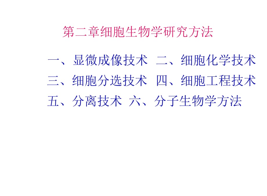
1、第二章细胞生物学研究方法一、显微成像技术 二、细胞化学技术 三、细胞分选技术 四、细胞工程技术 五、分离技术 六、分子生物学方法第一节显微成像技术1、光学显微镜11.1 普通复式光学显微镜技术(图)A原理Ill分辨率是指区分开两个质点间的最小距离幽分辨率 r = 0.61人/n sina最大分辨率蓝光入=450nm, sin70 =0.94Ar = 0.3gm(n=l)? r = 0.2 |im(n=1.5)分辨极限与放大率光镜样本制作:样品固定、包埋、切片、染色 1.2常用的光学显微镜.体视显微镜(图)相差显微镜和录相增差显微镜 倒置显微镜(图) 暗视野显微镜(图) 荧光显微镜目 激光共焦点
2、扫描显微镜技术回32、电子显微镜(图)2.1 电子显微镜的基本知识r 电镜与光镜的比较lai电镜的分辨率o. 2nm超微结构(亚显微结构)方电子显微镜的基本构造Iffll照明系统、成像系统、真空系统、记录系统国为什么电子显微镜需要真空系统2.2 透射电子显微镜(TEM)超薄切片技术(图):细胞超微结构的观察技术用于电镜观察的样本制备示意图Iffll与LM相比,EM样固定、包埋、切片、染色品组织固定有什么特殊要列,负染色技术(Negative staining)金属投影(图)染色背景,衬托出样品(病毒等)的精细结构(轮廓)、冰冻蚀刻技术(Freeze etching) (冰冻断裂、蚀刻、复型:作
3、用:主要用来观察膜断裂面的蛋白质颗粒和膜表面 结构。快速冷冻深度蚀刻技术IE17 电镜三维重构技术Iffll| 膜蛋白、病毒以及蛋白质一核酸复合物等大的复合体的三维结构。2.3 扫描电镜(SEM)原理与应用Iffll样品表面或断面的形貌特征的观察技术电子“探针”扫描,激发样品表面放出二次电子(图), 探测器收集二次电子成象。样品制备:取材,清洗,固定,脱水,干燥,粘样,喷金 环境扫描电镜显示生物样品的真实面貌 观察常规扫描电镜较难处理的样品生物样品的x射线元素分析生物样品的活体观察2.4 扫描遂道显微镜目第二节细胞化学技术,、酶细胞化学技术 酶细胞化学技术就是通过酶的特异细胞化学 反应来显示酶
4、在细胞内的定位。免疫细胞化学技术侬*免疫荧光技术:对抗原进行细胞内定位 圣免疫电镜技术:进行亚细胞结构和分子定位.第三节细胞分选技术,、流式细胞仪的基本原理(应用:对细胞或染色体进行分选,并对单个细 胞或其他生物微粒进行定量分析,包括测量 细胞的大小、形状、细胞DNA、RNA含量和 细胞总蛋白等。:、分选(图)第四节细胞工程技术细胞工程(cell engineering):利用细胞生物学的原 理和方法,结合工程学的技术手段,按照人们预先的 设计,有计划地改变或创造细胞遗传的技术。、细胞培养细胞培养(cell culture):在体外模拟体内的生理环 境,培养从机体中取出的细胞,并使之生存和生长
5、的 技术。1、动物细胞培养(图)2、植物组织培养(图):、细胞融合与单克隆抗体技术1、2、3、细胞融合幽单克隆抗体技术显微操作术(图)三、动物细胞核移植克隆技术(图)第五节分离技术1、离心分离技术用途:分离细胞器与生物大分子及其复合物A类型:差速离心:分离密度不同的细胞组分Iffll, 密度梯度离心:精细组分或生物大分子的分离维离子交换层 析的原理是 什么?2、层析分离技术 凝胶过滤层析 亲和层析回 离子交换层析13第六节分子生物学方法V1、基因工程技术(图)沏2、基因作图与人类基因组计划A 3、PCR技术4、选择性基因剔除与转基因鼠(图) 5、乳腺生物反应器技术(图)17TubeMirror
6、NosepieceObjectivesFine FocusStage Claps DiaphragmCompound Light MicroscopeDloplrUectianicBlovruiflensesSI,金 adjumenlsCondswrDaylight14Parts of a Nikon compound microscope.fHuminator LightBase(a) Principal parts arid lunciionsFine focusing knobOcular (eyepiece) Remagnifies the image formed by the ob
7、jective lensCondenser Focuses light through speciStags Holds (he - microscope slide in positionCoarse focusing knobOcular lensIlluminatorBase with source of illuminationBody tube Transmits、 the image from the objective lens to the ocularArm.Objective lenses Primary lenses that magnify the specimenDi
8、aphragm Controls the amount qI light entering the condenser(b) The path ol light (botlom io top)21Figure A-6 The Compound Light Microscope, (a) A compound light microscope. (b) The path of light through the compound microscope.图3-1显微镜成像原理目镜 视网AB:被检物体L1;物镜AN1:倒立实像L2: A2B2:放大的倒立虚像L3:眼球晶状体 A3B3:膜上正立像,
9、F2:分别是物镜和目镜的焦平面0.2 mm(200 pm)minimum resolvableby unaided eye2从mx10minimum resolvable by light microscopeSTmNVoH。20 nm0.2 nm图3 - 3相邻两衍射斑的三种情况minimum resolvableby electron microscope1 m = 103 mm =106|nm =10s nmmovomarM diMi嫡看国amspecimen nmbaddcHd inwfiMor wtin丁郭啊H D加ribbim MMSI;时 Wribbon af sections
10、on .58婚驯泌必班/ asPhase plateAnnular daphraynDiiracted *Qht (pnase allered Ly spgnen)Direct lght (phae unaltered by spec men)二 1J _5 sourceIrrege ptareFigure A-7 Optics of the Phase Contrast Microscope, 【he ccrtg- uratoi of the opiicaf elements and the paths of light rays through the phase-contrast mic
11、roscope. Hnk lines recresent light drftracted b/ tw jceciwxn. okJ bbckl res rctresoni direct ight.Figure A-8 Phase-Contrast Microscopy. A ptiase-comrast micrograph of epithelial cel:s. The cells were obsgM in ai unprocessed and unstd r(J sw Atich is a n叼or advant of phase-contras: meroserp/B背置像A被检物体
12、像图3-8相差镜检的光路示意图22 相差聚光器:向显微镜提供环状光源I 相差物镜:物镜的后焦点平面处装有相板, 分为两部分,一部分可通过直射光,为半透 明的环状,叫共机面,另一部分可通过衍射 光,叫补偿面。 相板上通常有吸收膜和相位膜,吸收膜吸收光线,降低光透过率;相位膜推迟相位。 相差滤色镜23应用:无色透明活体标本的细微结构,检查,鉴定活体细胞(培养细胞,分离细胞)4Transmitted Light Contrast ModesFigure 2Figure A-17 Computer-Enha need Digi怡I Video Microscopy.This series oi mic
13、rographs shows how computers can be used to enhance images obiained with light microscopy. In ihis example, an image of several microtubules. v-4iich aie too small to be seen with unenhanced light microscopy, are processed la make them visibfe in detail (a) The(d)image resulting from elecvonic contr
14、ast enhancement of the original image (which appeared io be empty*. (b) The background of ihie enhanced image in (a), which is then c) subtracied from image (al leaving only the microtubules. Id) The final, detailed image resulting from electronic averaging of the separate images pfocessed as shown
15、in a-c集光器载物自主要应用: 集光器与载 物台之间工 作距离较 高,可以放 置培养皿、物镜转换器主体培养瓶等容 器,直接对 培养的细胞 进行照明和26底座观察27a原理:丁道尔现象,微粒 对斜射光反射或衍射,增 大了人眼可见性。A主要应用:主要观察的是物体的轮廓,分辨不清内部的微细胞构造,适 合于观察活细胞内的细胞核、 线粒体、液体介质中的细菌和霉菌等。Radiolarian in Brightfiell and Darkfield Illumination.7Figure 2 暗场镜下染色和未染色标本 荧光显微镜技术(Fluorescence Microscopy)原理(图)应用(图)
16、“直接荧光标记技术 间接免疫荧光标记技术主要应用:研究细胞内物质的吸收、运输以 及化学物质的分布及定位等。31滤掉其他光,透过激发光激发光受反射、折标本受激发产生荧光向四周发散2滤掉激发光,透 过荧光宽谱线发射光(汞灯)ExciterFitterBarrierFilterFigure 3射或衍射向四周发散Innage planeVlSlDlC light -Barrier filterFigure A-12 Optics of the Fluorescence Microscope.Figure A-15 Fluorescence Microscopy. Cultured dog kidney
17、 epithelial cells stained with the fluorescent stain phalloidinf which binds to actn microfilaments.电子显微镜的基本构造电子枪阳极聚光镜样品室物镜中间镜投影傥观察窗,荧光屏SpecimenObjec?ive lensSpecimen stagefigure A-21 A Transmission Electron Microscope.(a) A photograph and (b) a schematic diagram of aEM.Cathode (tengsten filamerl)Fi
18、rst condenser lensSecond condenser lensElectron gun (irside Wchnelt cylinder, net shown)Condenserlens systemIntermediate lensPfojec:o lensViewing screenPhoiograpnic plaie (in camera chamber)39(a)(b)Figure 18.11 A comparison between the information contained in images taken by a light and electron mi
19、croscope at a comparable magnification of 4500 times actual size, (a) A photo of skeletal muscle tissue that had been embedded in plastic sectioned at 1 pm, and photographed with a light microscope under an oil immersion objective lens. (b) An adjacent section to that used for part a that was ait at
20、 0.025 pm and examined under the electron microscope at comparable magnification to that in a. The resulting image displays a one- to tvvo-hundred-fold increase in resolution. Note the difference in the details of the muscle myofibrils, mitochondria, and the red blood cell contained in the capillary
21、. Whereas the light microscope cannot provide any additional information to that of a, the electron microscope can provide much more information, producing images, for example, of the structure of the individual membranes within a small portion of one of the mitochondria (as in Figure 5.20). (Courte
22、sy of Douglas E. Kelly mid M. A. Cahill.)杨扬树脂物件的制行过覆三毒图缓冲液清洗镀酸后固定水清洗切取材料Uno理)戊一醛前冏定70七聚合乙群系列脱水环筑丙烷置换Spurr树脂浸透包埋材料修包埋块半薄切片贴片烤片1% TB0町(b)Figure 18.14 Examples of negatively stained and metal- shadowed specimens. Electron micrographs of a tobacco rattle virus after negative staining with potassium phos
23、photungstate (a) or shadow casting with chromium (b). (Courtesy of M. K. Corbett.)Examples of negatively stained and metal-shadowed specimens.Electron micrographs of a tobacco rattle virus after negative staining with potassium phosphotungstate(a) or shadow casting with chromium(b).SpecimenShadowing
24、 and replicatingReplica viewed inelectron microscope(b)Figure 18.16 Freeze-fracture replicas, (a Procedure for th。formation of freoze-fractiire replicas as described in the text. Freeze-etching is an optional step in which a thin layer of covering ice is evaporated to reveal additional information a
25、bout thp structure of the siirfaco of the fractured specimen. (b) Replica of a frceze-fracturcd onion root cell showing Ihe nuclear envelope (NE) with its pores (NP), the Golgi complex (G), a cytoplasmic vacuole (V)z and the coll wall (CW). (Courtesy of Linid Bm心“J#Figure 18.17 Deep etching. Electro
26、n micrograph of a ciliary axoneme from the protozoan Tetrnhi/rnena. The ax- onemes were fixed, frozen, and fractured, and the frmen water at the surface of the fractured block was evaporated away, leaving a portion of the axoneme standing out in re* lief, as visualized in this metal replica. The arr
27、ow indicates a distinct row of outer dynein arm&. (From Llrsula W. Goi)den&itgh and )ohn E, Heitorf J, Cell Biol. 95:8004982; by43Figure A-35 X-Ray Diffraction. X ray ditfraction cam M used to edec/af structure at near aiornc resolution. X ray$ diffracted by atoms in crystals or Sbsrs just as light
28、mves ere dittncted pinholes In most cases, the specimens of interest io biologists are proie fis cv nideic acids The spo:ific example illustrated in this figure t$ a DMA fiber The resu ting diffraction patterns 郡e recorded pbotograchxallyand can men te anMyjed mathematical?y to deduce the molecular
29、struchire This ptxxograph depicts the actual Xy* diffractpattern used by Jaras Watson and Francis CriSc to dedxe ihe molecular structure of double-stranded DNA A ccmpjtef graphic nxiel ot ONA double he6x.(2) The resulting Oiffracdonpattern is analyzedmjrheovrtical(j) X rays diffracted by ONA tibe 如
30、量子力学中的隧道效应(隧道电流)、原子间作用力、磁力、 摩擦力等,并在计算机显示出来,从而反映出样品表面形貌信息、电特性或磁特性等。(图)稣点:,1)可对晶体或非晶体成像,无需复杂计算,且分辨本领高。(侧分辨率& 为0.10.2nm,纵分辨率可达O.Olnm );辞可实时得到样品表面三维图象,可测量厚度信息;,3)可在真空、大气、液体等多种条件下工作;非破坏性测量。4)可连续成像,进行动态观察5途纳米生物学研究领域中的重要工具,在原子水平上揭示样本表面的结构。芦DDNA、RNA和蛋白质等生物大分子及生物膜、病毒等的结构。47Motion in z directionSurface to be
31、imagedImage built up by successive scansx and y scanning motionsScanning tipFigure A-33 Scanning Tunneling Electron Microscopy.48Figure 9.20 The role of microtubule-associated motors in vesicle and organelle transport. This cultured cell has Wn doubly labeled with antibodies against both kinesin yel
32、low) and tubulin (green). The anti-kinesin antibodies are seen to stain membrane-bound vesicles associated with the microtubular network (seen as orange color where the two fluorescent antibodies overlap). (From Scott T. Brady and K few Pfister, J. Cell Sci. Supplement 74, 1992; by permission of Com
33、pany of Biologists Ltd.)AntibodyProtein A0.5 pm9Antigen z (catalase)Fc domainPeroxisomes FIGURE 5-17 Localization of a specific protein by electnMi microscopy of antibody-stained samples. (al First the ent is allowed to interact with its specific antigen (e g. catalase! ma section of fixed tissue. T
34、hen the section is treated with a co of protein A from the bacterium Staphylococcus auteus and electron-dense 5-7 nm diameter gold particles. Binding of this complex to the common Fc domain of antibody molecules the target protein, catalase in this case, visible in the electron microscope, lb) A sic
35、e of liver tissue was fixed with glutara sectioned, and then treated as described in (a) to localize catalase. The gold particles (black dots) indicating the pre of catalase are located exclusively in peroxisomes. IFrom H. J. Geuze et a . 1981. J. Ceff Bro/. 89:653. Reproduced from the Cef) Bioiogy
36、by copwight permission of Th Rockefeller University PressProben strom返吊Y;*5;,加T6冷支号现.I*a51. cell suspension4nozzlelaser beam4. cell cylture, analysis, etc.flow cytometersort etectro-niCSEHwa什2000jan023. cell sorting2. staining ofmembrane,cytoplasm,and nucleus51细胞标记drop-chargingsignalsheath fluidnsio
37、nnonfluore-scenX cell collector fluorescent antibodies,Itrasomc nozzle vibrator3The dilute cell suspension contains tw。different types of cellswith differeni: surface antigens. The fluorescent antibodies arespecific for only one type of cell-surface antigen.So, on? cell type binds a label and the ot
38、her does not.染色体标记53drop-chargingsignal .-.i=-sheath fluidinterferenceultrasonic nozzlevibratorchromosome suspension-fluor esce-nV chromosome collectornonfluore-scenf- chromosomecollectorfl5sk for undeflected droplets0P K3Q Q Q J fluorescence-2000V-|H Q-+2000Vpoly nuckotide- probeswith fluorescentma
39、rkers attached55七a quxf3f:觉以P 3也加京路d 附而isw cyfme riu mZyx of* c PAII植物组织培养5657植物原生质体培养HETEROKARYONHYBRID CELL FIGURE 6-8 Fusion of cultured animal cells, (al Unfused growing mouse cells with a single nucleus per cell. (b) Fused mouse cells with 2-5 nuclei per cell. Fusion was induced by treatment wi
40、th polyethylene glycol (45 percent) for 1 minute. The number of nuclei per fused cell (heterokaryon) is determined by the polyethylene glycol concentration and the time of exposure. By adjustment of these factors, the number of heterokaryons containing only two nuclei can be maximized. From R. L. Da
41、vidson and R S. Gerald. 1976. Som. Cell Genet. 2 165.ifject mouse用3 Xmyoionna cc4i$ mble to grow m HATSome mouse lymphocytes E f-pccn make MMMXMDXMH“Ils ceCvXurv cells In separate weHsMu and fuse cel s. nwer to HAT nigunHytrktomasTest each gi 3 a-itibody g XFigure 18.45 The proccd une used in thanll
42、uiir- guamne phcTsphonbov I transtcraj*,HQlKli t(gnnv, but It $*、not support the grtnvth M cells kKkinc thi mrmc, uich as the urfuwd fnyclMiuedkuwd iMhkpwccOssocate ceK oi btestocyst nd cultireEScelsMonol w” noxJvidng cl$Transfect cels with DNA containing mutant gene XEScelsTransiected ES cell (hete
43、rozygous for mutant gene X)J L Grow celb m 7 selectivemedumHost is homozygous for the attxno al&e and lacks pigmenabonI=Inject translectedES cels intohost blastocystcontaining inner cel toss with two types of ceilsTo determm whetne* germ cels are denied from injected ES cels, mate the chmenc mou” wi
44、th ment)ef of aibino strainNonchmenc mouse, heterozygous fcr gene X.Knockout mice are pr(xkced by matng two of these heterozygotes61 shnercel mass (cooUns ES ceRs)Monolayef ofTropbectoderm cellsBUSlOCglTransfect cels with DNA containing mmant gene XTransfected ES cdl (heterozygous for mutant gene XI
45、EScekGrow 8ll$ in selective medum=Inject transtectedES cefs intohost blastocystHost is hocnozygevs for the albino altete and lacks pigrrentationOimeric mouse wth albnoand pigmented coatChimera bldstocyt ccntaining inner cel mass with two tytes of celtsNoochmenc mouse, heterozygous fcr gene X.Knockout mice are produced by mat ng two of these heterozygotes.To determine whether germ ceils are derrved fran injected ES cds. mate the chnwnc mouu 7 merrbef of aibino strainCells in arrested (Gq) phase
- 温馨提示:
1: 本站所有资源如无特殊说明,都需要本地电脑安装OFFICE2007和PDF阅读器。图纸软件为CAD,CAXA,PROE,UG,SolidWorks等.压缩文件请下载最新的WinRAR软件解压。
2: 本站的文档不包含任何第三方提供的附件图纸等,如果需要附件,请联系上传者。文件的所有权益归上传用户所有。
3.本站RAR压缩包中若带图纸,网页内容里面会有图纸预览,若没有图纸预览就没有图纸。
4. 未经权益所有人同意不得将文件中的内容挪作商业或盈利用途。
5. 装配图网仅提供信息存储空间,仅对用户上传内容的表现方式做保护处理,对用户上传分享的文档内容本身不做任何修改或编辑,并不能对任何下载内容负责。
6. 下载文件中如有侵权或不适当内容,请与我们联系,我们立即纠正。
7. 本站不保证下载资源的准确性、安全性和完整性, 同时也不承担用户因使用这些下载资源对自己和他人造成任何形式的伤害或损失。
